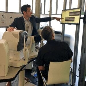
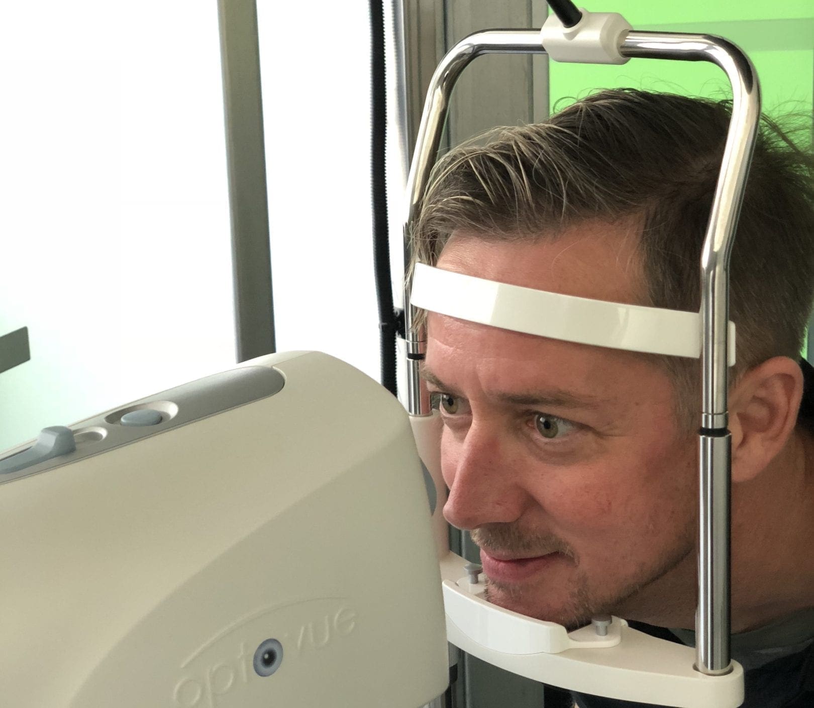
 At Eclectic Eye, we take advantage of modern innovations, technologies and screening devices. “OCT” stands for Optical Coherence Tomography, which essentially creates a map of the structures of your eye. We use OCT technology at the beginning of every comprehensive exam. This noninvasive scan uses infrared and long wavelength light to penetrate through the patient’s pupil to obtain 2D and 3D images and cross-sections of specific tissues. Over time, this device can track minuscule changes of specific tissues that your doctor may not be able to chart as accurately.
 This “optical ultrasound” has better resolution than other medical imaging such as MRIs and ultrasounds (which you can find more info about the latter here), because it uses light to gather data rather than radio frequencies or sound. OCTs have been used in other areas of the medical field like cardiology and dermatology and have even been commissioned in art conservation projects to analyze the different layers in paintings. The OCT scanner can aid our doctors in diagnosing various ocular conditions such as glaucoma, age related macular degeneration, diabetic eye disease and retinal issues.
There are numerous benefits of OCT imaging. There is no prep time for the patient, and the  doctor can obtain live, subsurface images at near microscopic resolution, sometimes even catching abnormal or damaged tissue before it affects the patient’s vision. The data collected can then provide tissue measurements, with size and depth to produce a 3D image of the patient’s tissue and optical structures almost immediately following the initial scan. Other benefits of the OCT machine, at this time, consist of no known side effects for the patient, no dilation is required, and the doctor can add the results to the patient’s record to compare year after year.
How an OCT Scan Works
 The process for the OCT scan is very simple. It starts with the technician or doctor sanitizing the machine for the patient, and then the patient is positioned in the device. The OCT tech or doctor then moves the imaging device closer to the patient’s eye, calibrates the machine and focuses on the tissue. The operator instructs the patient not to blink and asks them to focus on the target, and then three, two, one – the scan is complete without ever touching the patient’s eye. The doctor is able to get images of the patient’s retinas and retinal tissue, the optic nerve and blood vessels entering the optic nerve, corneal thicknesses and many other important optical structures and data.
The OCT helps the doctor with early detection and prevention of many ocular conditions and considerably improves treatment options for patients. As the field of imaging and optical devices continues to develop, Eclectic Eye, our doctors and opticians will be sure to stay up-to- date with the latest advancements in technology, ensuring you get the care and treatment you deserve. If you have any questions regarding the OCT process or would like to book a comprehensive exam, contact us today.


